OPHTHALMOLOGY TECHNOLOGIES
The Ellex Tango Reflex is a combined Selective Laser Trabeculoplasty (SLT) and Yttrium Aluminum Garnet (YAG) laser. It allows the ophthalmologists at Fredericksburg Eye Associates to perform four different procedures in clinic with the same laser:
-
YAG Capsulotomy for secondary cataract or posterior capsular opacification (PCO). After cataract surgery, the bag or capsule that holds the intraocular lens (IOL) will become cloudy or opacified about 10% of the time. This is a painless 60 second procedure that usually restores the vision to how it was right after cataract surgery.
-
YAG vitreolysis for symptomatic floaters. The Ellex Reflex laser is the first YAG laser to ever be used in a randomized controlled trial demonstrating the safety and efficacy of vaporizing floaters with a laser. Significant technological improvements were made over older lasers to improve the surgeon's view of floaters and allow for a safer, more effective procedure. Not every floater can be lasered safely and the doctors at Fredericksburg Eye Associates perform a thorough examination with specialized imaging to determine if a patient is a good candidate or not.
-
YAG iridotomy for narrow or closed angle glaucoma. The laser is used to create a tiny hole in the iris to prevent acute angle closure glaucoma, a dangerous and sight-threatening condition.
-
SLT for open angle glaucoma. SLT has been used for many years with success to lower patient's intraocular pressure. It is a safer alternative than more invasive glaucoma surgeries and can sometimes be used instead of glaucoma medications.
Alcon Centurion Phacoemulsification System


The Centurion Phaco System is the latest generation machine for cataract removal. It uses ultrasound energy to break up cataracts and aspirates them from the eye in tiny pieces. There are several improvements in this version compared to older technologies:
-
Less phacoemulsification energy is required to remove cataracts. This means the machine is gentler on the eye and has less difficulty removing extremely dense cataracts. Studies have shown improved results.
-
Active fluidics means fewer complications because the eye is less likely to collapse during surgery.
-
Since removal of the cataract is more efficient, this reduces the amount of time surgery takes.
-
The anterior vitrectomy cuts at a much faster rate than older machines--this is gentler and safer.
-
The ability to do a surgical iridotomy automatically without the use of scissors.
At Fredericksburg Eye Associates, we use phacoemulsification to remove cataracts and do not offer patients femtosecond laser cataract surgery. This is because using a femtosecond laser has not been proven to be safer or more accurate than phacoemulsification of cataracts. There are several randomized clinical trials comparing the two methods, but they all conclude they are equally safe and accurate. Use of the laser also usually increases costs and makes surgery take longer.
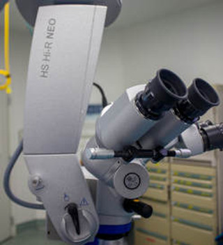
Haag Streit Hi-Resolution Neo 900 Microscope


The Haag Streit Hi-R Neo 900 is our operating microscope. We perform our surgeries at Hill Country Memorial Hospital with this high resolution, precision instrument. This is a significant upgrade over older technologies for the following reasons:
-
Improved resolution, focus, and view.
-
Electromagnetic controls and braking provide smoother surgeon control of the microscope.
-
Improved tilt of the microscope allows for better visualization of the intraocular angle. This improves the success in placing the iStent and other angle surgeries.
-
Integration with our Lenstar LS900 using a monitor attached to the operating microscope which displays the patient's name, ocular measurements, intraocular lens calculations, and lens alignment information. You can see a photo here.
-
The ability to record surgical video in high definition. If you want a recording of your surgery, just ask your surgeon BEFORE the surgery.

The Optos Monaco takes ultra wide field color and auto fluorescent photographs of the retina. It allows a view of the peripheral retina, even without dilation. It can be used to image a floater, retinal tear, retinal detachment, diabetic retinopathy, choroidal nevus, and other retinal pathology. It also performs macular OCT all on the same machine. Sometimes it is used to image the retina and dilation can be avoided but it depends on the patient and the clinical scenario. If you prefer retinal imaging to dilation, there may be an additional charge if not covered by your medical insurance. Ask your technician before dilation!
Haag Streit Lenstar LS900 APS

-
Optical measurement of ocular axial length is more accurate than older methods.
-
Proprietary 32-point, dual-zone keratometric data provides measurement of corneal power and astigmatism. This has been validated by research and found to be very accurate.
-
Measurement of anterior chamber depth and lens thickness to improve effective lens position calculations.
-
Use of 4th generation IOL formulas. We currently use the Barrett Universal II formula which has been compared to all other formulas and found to be the most accurate available.
-
Integration of the Lenstar data and photographs with our Haag Streit Hi-R Neo 900 Operating Microscope. This allows for confirmation of the correct lens power during cataract surgery and provides photographs for proper IOL alignment inside the eye
Haag Streit Slit Lamps

At Fredericksburg Eye Associates, all of the slit lamps we use are made by Haag Streit. The slit lamp is the device an eye doctor uses to examine your eyes in clinic. Haag Streit slit lamps are widely considered by ophthalmologists to be the best in visual quality, allowing accurate and efficient diagnosis of eye problems if present. We utilize our slit lamps as an indirect ophthalmoscope with a 90 or 66D lens to provide a binocular view of the retina and optic nerve. Two of our Haag Streit Slit Lamps are equipped with anterior segment cameras allowing high definition photographs and video of the front of the eye.
Humphrey Autorefractors

An autorefractor is a device that is used to determine a rough estimate of your glasses prescription. We have two Humphrey autorefractors which are integrated directly into our electronic medical record. Although the autorefractor can give a rough estimate of your glasses prescription, a refraction is required to determine your true glasses prescription, should you want new glasses or contacts.
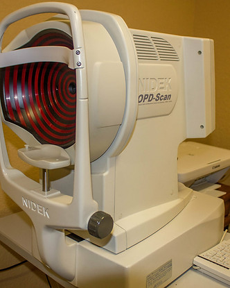
Nidek OPD Scan II Corneal Topographer

The Nidek OPD Scan II is our corneal topographer. It is used to create a map of the front surface of the cornea. This allows us to diagnose corneal ectasia such as keratoconus, pellucid marginal degeneration, and post-LASIK ectasia. It also allows for more accurate analysis of corneal astigmatism, including the amount, axis, and regularity of it. The corneal topographer is used in conjunction with the Lenstar LS900 for all patients desiring astigmatism correction at the time of cataract surgery. Since the Nidek OPD Scan II is a newer generation machine, it is able to provide intraocular aberrometry to assess for higher order aberrations which can cause visual complaints not correctable with glasses.
Optovue Avanti Widefield OCT

Ocular Coherence Tomography (OCT) uses light waves to produce high resolution images of ocular structures. It can be thought of as similar to Magnetic Resonance Imaging (MRI) or Computerized Tomography (CT) except that it is performed within minutes in an office setting without any kind of radiation or known side effects.
The Opotvue Avanti is a spectral-domain, high resolution OCT which has been utilized in numerous clinical trials. We use it to perform the following tests:
-
Nerve Fiber Layer and Ganglion Cell scans to screen for glaucoma and follow patients over time.
-
Retinal macular scans to diagnose and follow numerous medical eye problems including macular degeneration, diabetic macular edema, epiretinal membrane, vitreomacular traction, cystoid macular edema, etc.
-
Anterior Segment scans to diagnose corneal or iris pathology.
-
OCT Angiography which is starting to be used more than traditional fluorescein angiography to image the blood vessels of the retina and optic nerve. It is particularly helpful imaging patients with diabetes, macular degeneration, retinal vein occlusion, and glaucoma.
-
Measurement of Total Corneal Power (TCP) for calculation of Intraocular Lens (IOL) power after LASIK, PRK or RK which has been shown in studies to be the most accurate technology available.
-
Evaluation of the anterior chamber angle to determine the risk of acute angle closure glaucoma.
-
Corneal thickness mapping to judge central corneal thickness and screen for keratoconus.
-
Imaging of intraocular tumors such as a choroidal nevus or choroidal melanoma.

Humphrey Visual Field

The Humphrey Visual Field is our visual field machine. It is used most commonly to screen and follow glaucoma patients. The user sits in front of it and presses a button every time a small light is seen inside the machine. This machine is capable of testing the central and the peripheral vision. Although it is most commonly used to test glaucoma patients, it is also used to test patients for retinal toxicity from plaquenil (hydroxychloroquine), vision loss from a brain tumor, or vision loss following a stroke.


